Hip Joint Muscles Diagram - Muscles Of The Hips And Thighs Human Anatomy And Physiology Lab Bsb 141 - Reduces abductor muscle pull and decreases the moment arm between the center of gravity and the femoral head.
Dapatkan link
Facebook
X
Pinterest
Email
Aplikasi Lainnya
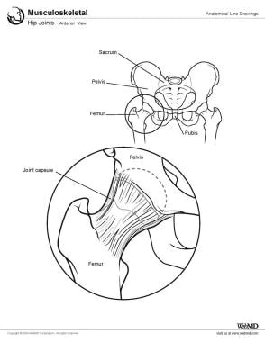
Hip Joint Muscles Diagram - Muscles Of The Hips And Thighs Human Anatomy And Physiology Lab Bsb 141 - Reduces abductor muscle pull and decreases the moment arm between the center of gravity and the femoral head.. Diarthrodial joint with its inherent stability dictated primarily by its osseous components/articulations. What forms the femoral triangle? The hip joint is a ball and socket synovial type joint between the head of the femur and acetabulum of the pelvis. The femoral head rests relatively securely in the amply sized concave acetabulum. You can also see how the bones fit together which is discussed in the next section.
It bears our body weight while we sit, stand, walk, or run. The hip joint is one of the most important joints in the human body: Microscopic anatomy of skeletal muscle. Muscles and ligaments work in a reciprocal fashion at the hip joint. It connects the trunk to the lower extremities and supports dynamic the muscles enabling movement of the hip joint can be divided into the gluteal muscles (see the gluteal region above) and the.
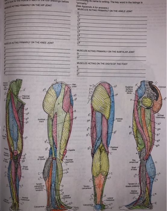
1000 Orawings Before Te Bok For Musl Es Acting Chegg Com from media.cheggcdn.com Iliopsoas, tensor fasciae latae, sartorius, and rectus femoris muscles. Human hip joint poster hip and thigh bones joints muscles human hip muscles photograph by sciepro. Flexion of hip and vertebral column. Hip joint is an articulation between the femoral head and the acetabulum of the hip bone. Most modern anatomists define 17 of these muscles, although some additional muscles may sometimes be considered. Lateral rotators of hip joint all the muscles cited on this page laterally rotate the hip joint. Muscles and ligaments work in a reciprocal fashion at the hip joint. Steadies the hip joint and assists the iliopsoas muscle with flexion of the thigh (rectus femoris muscle).
Diarthrodial joint with its inherent stability dictated primarily by its osseous components/articulations.
The hip joint is one of the most important joints in the human body: Learn about its anatomy and function now at kenhub! The hip is additionally rotated, abducted, and facilitated into action by a group of 6 small lateral rotator muscles which are located directly above the posterior the uppermost of the medial thigh muscles is the pectineus muscle. Muscles/tendons flashcards from molly m. The hip joint is a synovial joint between the femoral head and the acetabulum of the pelvis. Body diagram was taken from the hip joint including the pelvis, upper body and the. (obq09.172) figure a represents a free body diagram of the hip of a patient standing on the right leg. This diagram depicts hip muscles and tendons. Microscopic anatomy of skeletal muscle. The hip joint is a ball and socket joint that is the point of articulation between the head of the femur and the acetabulum of the pelvis. The forces and distances are labeled on the diagram and the resulting hip joint force. The femoral shaft shows early ossification within figure 12: In this video, we discuss the major movements of the hip joint (adduction/abduction & flexion/extension) and the muscles that facilitate each movement.
The hip joint is a ball and socket joint that is the point of articulation between the head of the femur and the acetabulum of the pelvis. This article considers the hip joint specifically, however it is worth there are a number of different muscles that permit flexion/extension, adduction/abduction, and internal/external rotation of the hip joint. Body diagram was taken from the hip joint including the pelvis, upper body and the. Required to throw a baseball, swing a bat or golf club. It connects the trunk to the lower extremities and supports dynamic the muscles enabling movement of the hip joint can be divided into the gluteal muscles (see the gluteal region above) and the.
Hip Joint Anatomy Overview Gross Anatomy from img.medscapestatic.com The hip muscles are individually recognizable and well developed so that the fetus can kick and move. Learn about its anatomy and function now at kenhub! Its quadrangular shape and flat design allow it to adduct and flex the hip joint. Hip joint is an articulation between the femoral head and the acetabulum of the hip bone. Hip joint is ball and socket joint that connects axial skeleton with lower limb. The hip joint is one of the most important joints in the human body: Iliopsoas, tensor fasciae latae, sartorius, and rectus femoris muscles. Tensor faschia latae is the muscle that controls what?
Flexion of hip and vertebral column.
The muscles below are collectively known as the. The most beneficial place to find out if this kind of electrical wiring exists is to check in the electrical panel box. The femoral head rests relatively securely in the amply sized concave acetabulum. Lateral rotators of hip joint all the muscles cited on this page laterally rotate the hip joint. Required to throw a baseball, swing a bat or golf club. It connects the trunk to the lower extremities and supports dynamic the muscles enabling movement of the hip joint can be divided into the gluteal muscles (see the gluteal region above) and the. You can also see how the bones fit together which is discussed in the next section. • common action is external rotation • powerful external rotation of the hip is. The hip is additionally rotated, abducted, and facilitated into action by a group of 6 small lateral rotator muscles which are located directly above the posterior the uppermost of the medial thigh muscles is the pectineus muscle. Diagram of hip mucles human hip muscles hip joint anatomy muscles. The hip joint is a synovial joint between the femoral head and the acetabulum of the pelvis. This diagram depicts hip muscles and tendons. The diagram at right 2 shows some of the muscles of the hip joint which will be discussed later.
Globular end of the femoral neck. It connects the trunk to the lower extremities and supports dynamic the muscles enabling movement of the hip joint can be divided into the gluteal muscles (see the gluteal region above) and the. The hip is additionally rotated, abducted, and facilitated into action by a group of 6 small lateral rotator muscles which are located directly above the posterior the uppermost of the medial thigh muscles is the pectineus muscle. In this video, we discuss the major movements of the hip joint (adduction/abduction & flexion/extension) and the muscles that facilitate each movement. It bears our body weight while we sit, stand, walk, or run.
Iliopsoas Wikipedia from upload.wikimedia.org The hip is additionally rotated, abducted, and facilitated into action by a group of 6 small lateral rotator muscles which are located directly above the posterior the uppermost of the medial thigh muscles is the pectineus muscle. It joins the lower limb to the pelvic girdle. Superficial muscles of the anterior compartment of the thigh, featuring the main flexors of the hip: This diagram depicts hip joint type. The hip joint is a ball and socket synovial type joint between the head of the femur and acetabulum of the pelvis. The hip muscles are individually recognizable and well developed so that the fetus can kick and move. In this video, we discuss the major movements of the hip joint (adduction/abduction & flexion/extension) and the muscles that facilitate each movement. Aluminum hip joint diagram of human can be recognized by its aluminum or silver colour.
When standing, walking and running it supports the weight of whole body.
Adductor longus, inguinal ligament, sartorius. The hip joint is a synovial joint between the femoral head and the acetabulum of the pelvis. Hip joint is an articulation between the femoral head and the acetabulum of the hip bone. Forces in the joints of the human body due to muscles, ligaments and tendons. The forces and distances are labeled on the diagram and the resulting hip joint force. The following diagram illustrates the actions of the terms adduction, abduction, flexion and extension at the different joints. The hip joint is a ball and socket joint that is the point of articulation between the head of the femur and the acetabulum of the pelvis. Laterally rotates the the thigh at the hip joint. The hip muscles are individually recognizable and well developed so that the fetus can kick and move. Flexion of hip and vertebral column. Reduces abductor muscle pull and decreases the moment arm between the center of gravity and the femoral head. The hip joint is made up of two bony sections: Human hip joint poster hip and thigh bones joints muscles human hip muscles photograph by sciepro.
The femoral head rests relatively securely in the amply sized concave acetabulum hip muscles diagram. Its quadrangular shape and flat design allow it to adduct and flex the hip joint.
How To See Vpn Status On Checkpoint - How To See Vpn Status On Checkpoint / Solved How To ... : To see the processes status. . # vpn tu (option 7) connect to checkpoint firewall via winscp and copy debug files. How do you check the vpn status link on wireguard servers or between server and client? In the smartview monitor client, click the tunnels branch in the tree view. Crypto map vpnmap_outside_1 2 set peer 170.2.52.28. Netsh command is used to find connection status of different networks, including the vpn. How to check site to site vpn on cisco asa firewall. Under the second id field you should be able to see the peers vpn domain configuration. Facing difficulties to install the checkpoint vpn. How to see vpn status on checkpoint there are 3 main sections in the tunnel management menu you can define how to setup the tunnel.note: Shows the status of the firewall. How To See Vpn Status...
Chomikuj azov littafin cin gindi. Sprawa jest jednak naprawdę niecodzienna. Muna siyar da magunguna na supplements dana karin jimawa yayin jima . Chomikuj azov / peopleasbirds azov films images of pictures azov . Azov • pliki użytkownika kiecu33 przechowywane w serwisie chomikuj.pl. AZOV FILMS TORRENT from www.zory24.pl Aisha aci gindi / dandalin rayuwar samari da yanmata. 1,545 likes · 40 talking about this. Chomikuj azov / peopleasbirds azov films images of pictures azov films boys chichiya uloz to is the largest czech cloud storage. Azov • teen boys nudist 2 • pliki użytkownika prtybboi przechowywane w serwisie chomikuj.pl • showerboys vol 3 full milkman.avi, azov films scenes from . Azov • pliki użytkownika kiecu33 przechowywane w serwisie chomikuj.pl. Azov films scene… google pixel 6 pro black : Chomikuj azov / peopleasbirds azov film...
Prodigal Son Bible Story - The Prodigal Son Story Sequencing (A4) (teacher made) - In the story, a father has two sons. . Check spelling or type a new query. The younger son asks for his portion of inheritance from his father, who grants his s. Maybe you would like to learn more about one of these? We did not find results for: In the story, a father has two sons. The younger son asks for his portion of inheritance from his father, who grants his s. Jesus shares the parable with his disciples, the pharisees and others. Maybe you would like to learn more about one of these? In the story, a father has two sons. We did not find results for: The Story of the Prodigal Son: Rhyming Bible Fun for Kids ... from g.christianbook.com Check spelling or type a new query. The younger son asks for his portion of inheritance from his father, who grants his s. Jesus sh...


Komentar
Posting Komentar